Research
Please visit the above links to have a more detailed look at our research projects. Below is a list of Centers that we are leading or affiliated with.
NSF MBM at UIUC - Miniature Brain Machinery
NSF STC EBICS at MIT/GT/UIUC - Emergent Behavior of Integrated Cellular Systems
NSF IGERT at UIUC - Cellular and Molecular Mechanics and Bionanotechnology
(download brochure)NIH Training Grant at UIUC - Midwestern Cancer Nanotechnology Training Center
(download brochure)NSF CiiT (I/UCRC) at UIUC - Center for Innovative Instrumentation Technology
NSF NSEC at OSU - Center for Affordable Nanoengineering for Polymeric Micro and Nanodevices
Research: >> Current >> Micro & Nanomechanical...
Micro and Nanomechanical Cantilever Sensors for biochemical Detection:
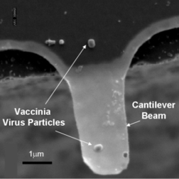
1. Micro-mechanical Sensors for Virus Detection
Amit Gupta, Dr. Demir Akin, School of Electrical and Computer Engineering
Prof. Steve Broyles, Department of Biochemisry,
Prof. Mike Ladisch, Department of Ag and Bio Eng/BioMedical Engineering
Prof. Rashid Bashir, School of Electrical and Computer Engineering/Department of Biomedical Engr.
Purdue University, West Lafayette, Indiana 47907
This research is funded by NIH
The long-term objective of this application is to develop a micro-scale, robust, real-time monitoring device, based on micro-machined ultrathin cantilever arrays for the rapid and sensitive detection of infectious agents, particularly bioterrorism agents in field setting and in primary-patient care facilities. The array will be specific for specific pathogens and will have the sensitivity to detect a single virus or toxin molecule. During Phase I, the proposed effort aims to develop dielectrophoresis-based infectious agent trapping, separation and concentration device and a proof-of-principle demonstration for the detection of an air-borne virus on functionalized micro-scale cantilever. The performance value of the devices for trapping, separation, concentration and detection of aerosolized coronavirus particles will be assessed. During Phase II, this sensor design and manufacturing capabilities will be extended and scaled-up to other infectious agents in the form of integrated sensor arrays with capability for on-board signal processing.
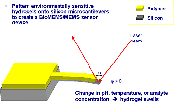
2. Environmentally Sensitive Hydrogels Patterned on Silicon Micro-cantilevers
Zack Hilt*, Amit Gupta, Prof. Nick Peppas*, Prof. Rashid Bashir,
School of Electrical and Computer Engineering, Purdue University
*School of Chemical Engineering
UT Austin, Austin, TX
Nature Materials Highlights on Swell Sensors
A process has been developed for patterning thin films of environmentally sensitive polymers on silicon microcantilevers. Intensive research has been focused on utilizing microcantilevers as highly sensitive chemical, physical, and biological microsensors. Hydrogels can be made sensitive to environmental conditions through the choice of functional groups in their polymer network. They are sensitive to temperature, pH, magnetic field, electric field, and ionic strength. By patterning these environmentally sensitive hydrogels onto silicon microcantilevers, microsensors can be prepared for MEMS or BioMEMS applications. We studied a cross-linked poly(methacrylic acid) containing large amounts of poly(ethylene glycol) dimethacrylate. This gel exhibits a swelling dependence on pH. By increasing the environmental pH above the pKa of poly(methacrylic acid) to cause ionization of the carboxylic acid groups, electrostatic repulsion is produced along the main polymer chains causing the polymer network to expand and swell. Therefore, a pH change induces swelling or shrinking of the polymer network and create stress on the microcantilever surface causing it to bend. In this study, silicon microcantilevers were fabricated on p-type (100) SOI wafers. Covalent adhesion was gained between the polymer and the silicon surface, by modification of the silicon surface with g-methacryloxypropyl trimethoxysilane. Microcantilevers were fabricated with various lengths, widths, and thicknesses. Polymers were patterned onto the silicon microcantilevers utilizing standard photolithography techniques. A Karl Suss MJB3 mask aligner was utilized to achieve alignment with 0.1 mm precision. Images of the patterned hydrogels were obtained using optical microscopy and scanning electron microscopy. The bending of the microcantilever was observed optically utilizing an optical microscope.
This research is funded by NSF Grants DGE-99-72770 and ECS-99-84199.
References:
[1] J. Ward, R. Bashir, and N. Peppes, “Micropatterning of Biomedical Polymer Surfaces by Novel UV Polymerization Techniques”, Journal of Biomedical Materials Research, Volume 56, Issue: 3, 24th Apr 2001, Pages: 351-360
[2] R. Bashir, J.Z. Hilt, A. Gupta, O. Elibol, and N.A. Peppas, “Micro-mechanical Cantilever as an Ultra-Sensitive pH Micro-sensor”, Applied Physics Letters, Vol. 81, No. 16, pp 3091-3093, Oct 14th, 2002.
[3] J. Zachary Hilt, Amit Gupta, Rashid Bashir, and Nicholas A. Peppas, “Ultra-sensitive biomems sensors based on Microcantilevers patterned with environmentally responsive hydrogels”, Biomedical Microdevices, Volume 5, Issue 3. pp. 177-184, September 2003
3. Micro-mechanical Sensors for Biological Detection
Angelica Davila, Dr. Demir Akin, Amit Gupta, School of Electrical and Computer Engineering
Dr. Tom Walter, Prof. Arthur Aronson, Department of Biological Sciences,
Prof. Rashid Bashir, School of Electrical and Computer Engineering/Department of Biomedical Engr.
Purdue University, West Lafayette, Indiana 47907
The purpose of this application is to develop a micro-scale, robust, real-time monitoring device, based on micro-machined ultrathin cantilever arrays for the rapid and sensitive detection of infectious agents, particularly bioterrorism agents in field setting and in primary-patient care facilities. These cantilever arrays will be specific to Bacillus anthracis. The proposed experiment consists of applying the spores onto the cantilevers, germinating the spores and being able to detect the added mass of the bacteria. In order to detect the mass, the frequency of the cantilever will be measured before and after the application and germination of the spores. Furthermore, an enzyme specific to the bacteria will be used to lyse it on the cantilever and that mass will also be detected by the shift in frequency of the cantilever.
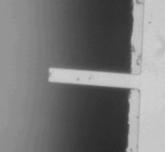 Figure 1: Bacillus anthracis spore on cantilever |
 Figure 2: Bacillus anthracis germinated spores |
Project Funded by NIH Grant and NASA INAC at Purdue
References:
[1] A. Gupta, J. Denton, H. McNally, and R. Bashir, “Novel Fabrication Method for Surface Micro-Machined Thin Single-Crystal Silicon Cantilever Beams”, Journal of Microelectromechanical Systems, Vl. 12, No. 2, April 2003, pp. 185-192.
[2] A. Gupta, D. Akin, R. Bashir, “Single virus particle mass detection using microresonators with nanoscale thickness”, Vol. 84, No. 10, pp. 8th March 2004