Research
Please visit the above links to have a more detailed look at our research projects. Below is a list of Centers that we are leading or affiliated with.
NSF MBM at UIUC - Miniature Brain Machinery
NSF STC EBICS at MIT/GT/UIUC - Emergent Behavior of Integrated Cellular Systems
NSF IGERT at UIUC - Cellular and Molecular Mechanics and Bionanotechnology
(download brochure)NIH Training Grant at UIUC - Midwestern Cancer Nanotechnology Training Center
(download brochure)NSF CiiT (I/UCRC) at UIUC - Center for Innovative Instrumentation Technology
NSF NSEC at OSU - Center for Affordable Nanoengineering for Polymeric Micro and Nanodevices
Research: >> Past >> Basic-Bio Inspired Assembly
Micro-fluidic Bio-chips for the Detection of Microorganisms
1. Microfluidic BioChips for the Electronic Detection of Bacterial Metabolism
Haibo Li, Yi-Shao Liu, Kidong Park, Rashid Bashir, School of Electrical and
Computer Engineering
T. Huang, M. Ladisch, Department of Agricultural and Biological Engineering
T. Geng, A. Bhunia, Department of Food Sciences
Purdue University, West Lafayette, Indiana 47907
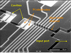
The development of techniques to process and detect bio-species using micro-fabricated systems will accelerate the practical applications of chip-based assays, with the ultimate goal of carrying out all of the operations on a chip, using minute sample volumes. In the first project described, the design and fabrication of micro-fluidic chips is being studied for affinity-based detection of biological species. The chip consists of a series of cavities or wells, which are anisotropically etched in a silicon wafer, connected by etched channels and sealed with a glass cover. The cavities have sizes that range from 80umx 80um to 530umx 850um and the channels have widths between 20um and 100um, which are all etched to a depth of 10um. Platinum electrodes are defined at the bottom of the cavities, whose surface is oxidized, and serve as the primary sensing and reaction elements when coated with receptors, antibodies, enzymes, etc. A spin-on-glass is used as an intermediate layer to bond the glass cover to the oxide-covered chip. The total fluidic path volume in the device is on the order of 30nl. Flow fields in the closed chip were mapped by particle image velocimetry. Electrical impedance measurements of suspensions of the live microorganism, Listeria innocua, injected into the chip demonstrate an easy method for detecting the viability of a few bacterial cells. By-products of the bacterial metabolism modify the ionic strength of a low conductivity suspension medium, significantly altering its electrical characteristics.
The work is now directed towards a recently funded project by USDA aimed at detection of Pathogenic Listeria in food products.
 |
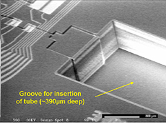 |
SEM Micrographs of micro-fluidic biochips |
|
References:
[1] A. K. Bhunia, Z. W. Jaradat, K. Naschansky, M. Shroyer, M. Morgan, R. Gomez, R. Bashir, and M. Ladisch, "Impedance spectroscopy and biochip sensor for detection of Listeria monocytogenes", SPIE Proceedings, Vol 4206-05, Nov 5-8, Boston, Mass. 2000.
[2] R. Bashir, R. Gomez, A. Sarikaya, M. Ladisch, J. Sturgis, and J. P. Robinson, “Adsorption of Avidin on Micro-Fabricated Surfaces for Protein Biochip Applications”, Biotechnology and Bioengineering, Volume 73, Issue 4, 2001, pp. 324-328.
[3] R. Gomez, R. Bashir, T. Geng, A. Bhunia, M. Ladisch, H. Apple, S. Wereley, “Micro-Fluidic Bochip for Impedance Spectroscopy of Biological Species”, Biomedical Micro-Devices, vol. 3, no. 3, September 14th, 2001, p. 201-209.
[4] Rafael Gomez, Rashid Bashir, Arun K. Bhunia, Michael R. Ladisch, “Microfabricated device for impedance-based detection of bacterial metabolism”, Spring MRS 2002. San Francisco, CA.
[5] Rafael Gómez, Rashid Bashir, Arun K. Bhunia, "Microscale Electronic Detection of Bacterial Metabolism", Sensors & Actuators B-Chemical. v 86 n 2-3 Sep 20 2002. p 198-208
Funded by USDA through the Food Safety Engineering Center at Purdue.
project web page at the center site.
2. Dielectrophoretic Characterization and Separation of Microorganisms in Micro-fabricated Device with Inter-digitated Electrodes
Haibo Li, D. Akin, Rashid Bashir, School of Electrical and Computer Engineering
Key Words: electrophoresis; dielectrophoresis; cell separation; micro-interdigitated electrodes; Listeria; BioMEMS.
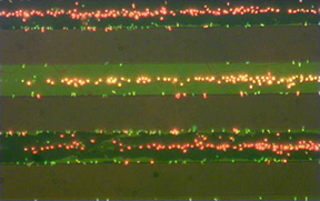
This work focuses on the electrophoretic and dielectrophoretic separation of live and heat-killed Listeria innocua cells on a microfabricated device with interdigitated electrodes. ANSYS and Mathematica are being used to perform the simulation of the electrical field and the dielectrophoretic force between the electrodes numerically and analytically respectively. Live and dead bacteria cells are stained with fluorescent dye (Live/DeadÒ BacLightTM Bacterial Viability Kit from Molecular Probes Corp.) and their electrophoretic and dielectrophoretic behaviors are observed under fluorescence microscope. Both DC and AC signals are applied to the intedigitated electrodes in the experiments. The frequency dependence of the applied AC voltage on the dielectrophoresis of the cells is studied with particular interest. Preliminarily results show the feasibility of using these methods to separate live and dead cells, as well as different microorganisms according to the different frequency dependences of their dielectric properties.
 |
|
Optical picture of interdigitated electrodes. Live and dead cells are separated at Vpp=1V, Frequency=50KHz, in DI water, most live cells (green) collect at the edges of the electrodes (positive DEP), while most dead cells (red) collect at the centers (negative DEP) |
Funded by USDA
[1] H. Li, R. Bashir, “Dielectrophoretic Separation Of Live And Heat-Treated Cells Of Listeria On Microfabricated Devices With Interdigitated Electrodes”, Spring MRS 2002. San Francisco, CA.
[2] Haibo Li, Rashid Bashir, “Dielectrophoretic separation and manipulation of live and heat-treated cells of Listeria on microfabricated devices with interdigitated electrodes”, Sensors & Actuators B-Chemical. v 86 n 2-3 Sep 20 2002. p 215-221