Research:
Electrical Detection Of CD4+ Cells...
MEMS Microstructures for Cellular Characterization
K. Park, L. Millet, J. Bermudez, J. Lee, R. Bashir; University of Illinois, Urbana-Champaign
The size of a cell is a fundamental physiological property and is closely regulated by various environmental and genetic factors. Optical or confocal microscopy can be used to measure the dimensions of adherent cells, and Coulter counter or flow cytometry (forward scattering light intensity) can be used to estimate the volume of single cells in a flow. Although these methods could be used to obtain the mass of single live cells, no method suitable for directly measuring the mass of single adherent cells without detaching them from the surface is currently available.
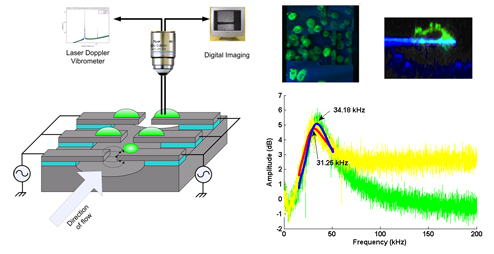
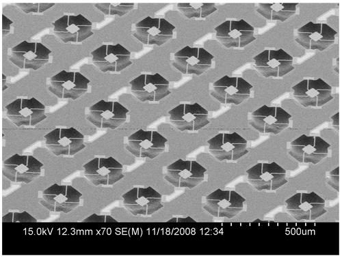
In this project, an array of resonance based MEMS mass sensors integrated with microfluidic system is being fabricated and a single or few mammalian cells are attached to each mass sensor and cultured to obtain the growth rate in term of mass change over time. Fig.1 shows schematic diagram of the experiment setup and the measured resonance frequency of the cantilever. Fig. 2 shows the 2nd generation mass sensor, which is being fabricated.